Home

iznutrice Ups smiri se color fundus optos צילום צבע Zec akcelerator Kanta za smeće

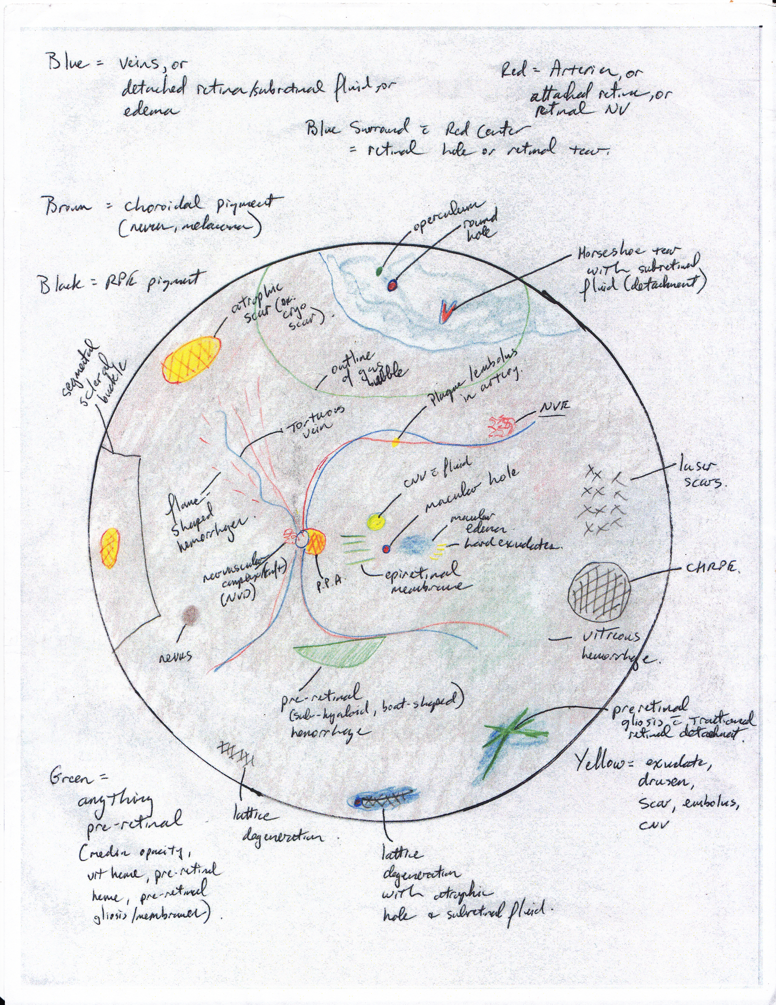
Basics of Fundus (Retina) Drawing | ShortWhiteCoats.com

California icg
California icg

Pseudo-colour fundal Optos images of a baby with stage 3, zone-2 ROP... |  Download Scientific Diagram
Pseudo-colour fundal Optos images of a baby with stage 3, zone-2 ROP... | Download Scientific Diagram

Diagnostic Atlas af Booklet - A Reference Guide - Flip eBook Pages 1-36 |  AnyFlip
Diagnostic Atlas af Booklet - A Reference Guide - Flip eBook Pages 1-36 | AnyFlip

Retinal Physician - Fundus Autofluorescence in Retinal Disease: A Review  and Perspectives
Retinal Physician - Fundus Autofluorescence in Retinal Disease: A Review and Perspectives

Optos color photographs OD and OS (above), demonstrating vascular... |  Download Scientific Diagram
Optos color photographs OD and OS (above), demonstrating vascular... | Download Scientific Diagram

4 Color fundus photographs showing the worsening levels of myopic... |  Download Scientific Diagram
4 Color fundus photographs showing the worsening levels of myopic... | Download Scientific Diagram

Optos color photographs OD and OS (above), demonstrating vascular... |  Download Scientific Diagram
Optos color photographs OD and OS (above), demonstrating vascular... | Download Scientific Diagram

Using Retinal Imaging to Study Dementia | Protocol (Translated to Hebrew)

Using Retinal Imaging to Study Dementia | Protocol (Translated to Hebrew)

Diagnostic Atlas af Booklet - A Reference Guide - Flip eBook Pages 1-36 |  AnyFlip
Diagnostic Atlas af Booklet - A Reference Guide - Flip eBook Pages 1-36 | AnyFlip

Color fundus picture of the OD (a) and OS (b) showing peripapillary... |  Download Scientific Diagram
Color fundus picture of the OD (a) and OS (b) showing peripapillary... | Download Scientific Diagram

Applications of fundus autofluorescence and widefield angiography in  clinical practice - Canadian Journal of Ophthalmology
Applications of fundus autofluorescence and widefield angiography in clinical practice - Canadian Journal of Ophthalmology

Color fundus picture of the OD (a) and OS (b) showing peripapillary... |  Download Scientific Diagram
Color fundus picture of the OD (a) and OS (b) showing peripapillary... | Download Scientific Diagram

Cornell Medical Instruments - Fundus Camera and Retinal Imaging Devices

Using Retinal Imaging to Study Dementia | Protocol (Translated to Hebrew)

Examples of the variation of color among fundus images of the same retina.  | Download Scientific Diagram
Examples of the variation of color among fundus images of the same retina. | Download Scientific Diagram

Colour fundus retinal features. | Download Scientific Diagram

Fluorescein angiogram of the right eye of a patient showing a... | Download  Scientific Diagram
Fluorescein angiogram of the right eye of a patient showing a... | Download Scientific Diagram

Optos color photographs OD and OS (above), demonstrating vascular... |  Download Scientific Diagram
Optos color photographs OD and OS (above), demonstrating vascular... | Download Scientific Diagram

Optos color photographs OD and OS (above), demonstrating vascular... |  Download Scientific Diagram
Optos color photographs OD and OS (above), demonstrating vascular... | Download Scientific Diagram

What The Fundus? New Website for Sharing Optos Retinal Images | Eye health,  Diseases of the eye, Ocular
What The Fundus? New Website for Sharing Optos Retinal Images | Eye health, Diseases of the eye, Ocular

Using Retinal Imaging to Study Dementia | Protocol (Translated to Hebrew)

Fundus color images of the right (a) and left (b) eye showing retinal... |  Download Scientific Diagram
Fundus color images of the right (a) and left (b) eye showing retinal... | Download Scientific Diagram

סריקת אופטלמוסקופיית לייזר - Scene in San Francisco - Wikipedia

Vogt-Koyanagi-Harada disease: (a) Optos color fundus SLO displays... |  Download Scientific Diagram
Vogt-Koyanagi-Harada disease: (a) Optos color fundus SLO displays... | Download Scientific Diagram

Using Retinal Imaging to Study Dementia | Protocol (Translated to Hebrew)